Design and Validation of a Robotic Knee Exoskeleton with Musculoskeletal Simulation-Derived Biological Torque Control

Rajiv V. Joshi1, Jimin An1, Ilseung Park1, Dawit Lee2, Inseung Kang1
1Carnegie Mellon University 2Stanford University

Abstract

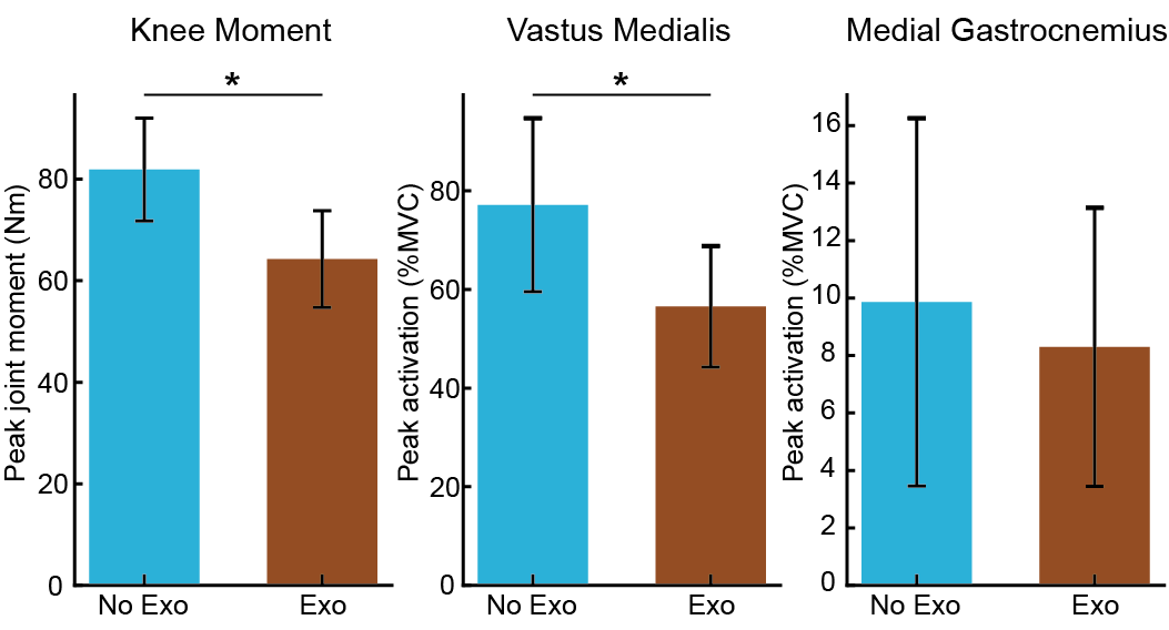
Data-driven controllers have enabled lower-limb robotic exoskeletons to adapt assistance to the user across many tasks. A leading example is biological torque control, which estimates the user's own joint moment from onboard sensors and assists in proportion to that estimate. However, its estimators are trained on data recorded with the target device, so deploying the controller on new hardware requires collecting new data. Existing open-source efforts release hardware, software, or controllers separately, so this data collection must still be repeated on the assembled device. Here, we present an open-source knee exoskeleton, built from off-the-shelf components and widely available fabrication tools, with a biological torque controller derived entirely from simulation. A muscle-driven imitation policy reproduces squatting motions from an open biomechanics dataset, and the resulting knee moments are distilled into an estimator that uses only onboard sensors, so no training data are recorded on the device. In seven able-bodied participants, assistance reduced peak biological knee extension moment by 21.5% and peak vastus medialis activation by 26.7% during squatting, relative to squatting without the exoskeleton (p<0.05). Our results demonstrate that a biological torque controller derived from simulation can transfer to physical hardware without device-specific data collection. Because the hardware and training pipeline are released together, research groups can train and compare knee exoskeleton controllers on shared hardware without additional data collection overhead.


Robotic Knee Exoskeleton Design

Overview of the robotic knee exoskeleton design
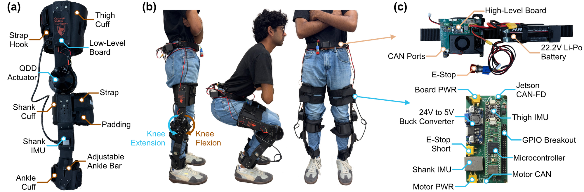
Figure 1. Overview of the robotic knee exoskeleton design. (a) Sagittal-plane view of the unilateral device, with straps, hooks, and cuffs at the thigh, shank, and ankle. (b) A participant wearing the bilateral device, shown left to right in sagittal standing, sagittal squatting, and frontal standing. (c) The high-level control board built around the embedded computer (top) and the low-level control board built around the microcontroller (bottom).

Table I. Device Specifications

Mechanical
Total mass, bilateral* (kg)3.5
Unilateral mass (kg)1.3
Actuator mass (kg)0.49
Rated / peak torque** (Nm)9 / 22
Range of motion (deg)0–120
Compute/Electrical
Inertial sensor communicationSPI
Motor communicationCAN
Low-to-High level communicationCANFD
Low-level loop execution (µs)~80
High/low-level round trip (ms)<0.5
Estimated run time (h)~2

*Includes the battery and embedded electronics.
**The stock motor driver limits output to 18 Nm per the manufacturer's safety constraint.

Simulation-Derived Biological Torque Control

Overview of the simulation-derived biological torque controller
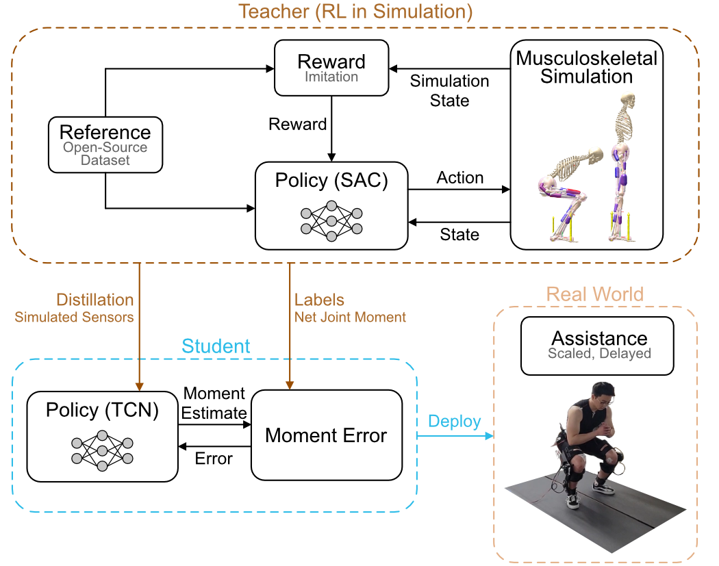
Figure 2. Overview of the simulation-derived biological torque controller. An imitation policy is trained with reinforcement learning in a musculoskeletal simulation to reproduce reference squatting motion, using an imitation reward as its learning signal. Knee muscle moments from the policy rollouts are then distilled into a temporal convolutional network that estimates the knee moment from onboard sensor signals, trained by minimizing the error against the simulated moments. On the exoskeleton, the estimated knee moment is scaled and delayed to generate assistive torque.

Experimental Validation

Experimental setup for validating the integrated exoskeleton and controller during squatting
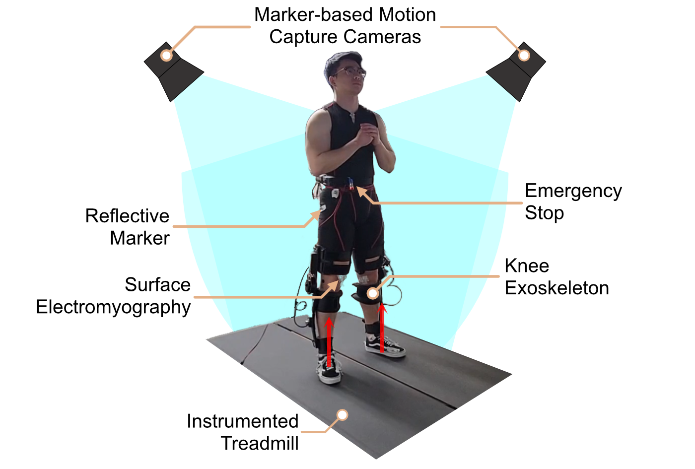
Figure 3. Experimental setup for validating the integrated exoskeleton and controller during squatting. A participant wearing the exoskeleton on the split-belt instrumented treadmill, whose embedded force plates measure ground reaction forces, with example placements of the electromyography sensors and reflective motion capture markers.

Results

Group-averaged knee kinematics, kinetics, and muscle activation during squatting
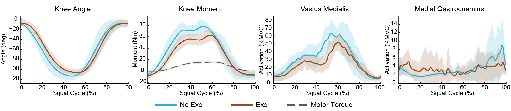
Figure 4. Group-averaged knee kinematics, kinetics, and muscle activation during squatting. (a) Knee angle, (b) biological knee extension moment with commanded torque, (c) vastus medialis activation, and (d) medial gastrocnemius activation over the normalized squat cycle. Solid curves represent group means across participants, and shaded regions represent ±1 standard deviation. Blue and brown indicate squatting without (No Exo) and with (Exo) the exoskeleton, respectively, and the dashed gray curve indicates the commanded torque with the exoskeleton. Muscle activation is expressed as a percentage of each participant's maximum voluntary contraction (%MVC).
Peak biological knee extension moment and muscle activation with and without the exoskeleton
Figure 5. Peak biological knee extension moment and muscle activation with and without the exoskeleton. (a) Peak biological knee extension moment, (b) peak vastus medialis activation, and (c) peak medial gastrocnemius activation without (No Exo) and with (Exo) the exoskeleton. Bars represent group means, and error bars represent ±1 standard deviation across participants. Asterisks indicate a significant difference between conditions (p<0.05, paired t-test).

Table II. Comparison with Existing Knee Exoskeleton Platforms

DeviceValidation SubjectsDevice Mass (kg)Peak Torque (Nm)TasksReported Reduction
This Study7 able-bodied3.518Squatting26.7% vastus medialis activation
Nesler et al.1 able-bodied1.6a30Lifting and lowering78.9% vastus medialis activation
Lee et al.10 able-bodied4.415Level walking, ramps, and stairs6.2% metabolic costc
Zhang et al.6 able-bodied3.218Level walking, ramp and stair ascent4.3–12.0% metabolic cost
Boon et al.8 able-bodied2.6a55Stair ascent26% vastus medialis activation
Chen et al.2 able-bodied4.135Kneeling18.4% vastus medialis activationb,c
Huang et al.8 able-bodied3.514Level walking19.6% vastus medialis activationc
Sarkisian et al.1 post-stroke3.6a40Sit-to-stand29.3% vastus medialis activation
Yu et al.7 able-bodied3.554Squatting38.1% vastus medialis activation,
12.8% metabolic cost

Mass is for a bilateral device unless noted. Activation reductions are in peak values and relative to no exoskeleton unless noted. aUnilateral device. bMean rather than peak activation. cRelative to the unpowered or zero-torque exoskeleton.

BibTeX

@article{joshi2026KneeExo,
  title   = {Design and Validation of a Robotic Knee Exoskeleton with Musculoskeletal Simulation-Derived Biological Torque Control},
  author  = {Joshi, Rajiv V. and An, Jimin and Park, Ilseung and Lee, Dawit and Kang, Inseung},
  journal = {arXiv preprint arXiv:2610.41981},
  year    = {2026}
}